Cookstown Dental Centre
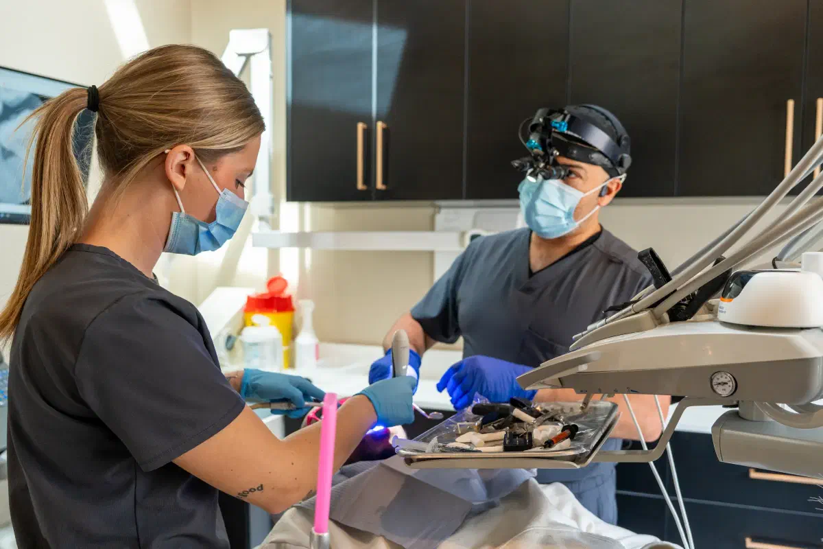
The Cookstown Dental Centre team caring for a patient in a treatment room

General & Family Dentistry · Cookstown

See the area your dentist is talking about.

Explore how close-up dental photographs can support explanations, document visible findings, and help you ask informed questions about your care.

Request an appointment

Let’s find a time that works.

Need urgent dental help? Call (705) 291-2222.

Intraoral Camera Examinations in Cookstown

A clearer conversation, one image at a time.

It can be difficult to understand a dental finding when you cannot see the back of your own mouth. An intraoral camera is a small camera used to capture close-up images of visible tooth and tissue surfaces. Seeing the area on a screen can help you follow an explanation and understand which tooth or restoration is being discussed.

At Cookstown Dental Centre, ask whether camera images would be useful for the concern being assessed. Photography can support communication and documentation, but an enlarged image is not a diagnosis by itself. The dentist combines what can be seen with your symptoms, examination, and other appropriate records before explaining the available options.

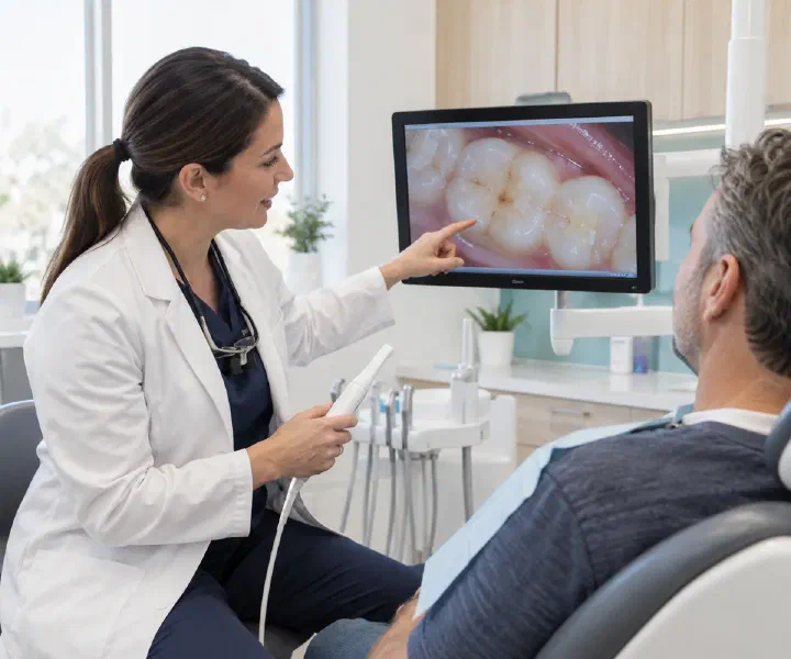
A dentist showing a patient a close-up photograph of molars

A closer look

Look, understand, and ask.

The value of a photograph is in the explanation that accompanies it. You do not need to interpret every colour or line on the screen to take part in decisions.

  1. Locate the concern

    The dentist can use an image to identify the tooth, surface, or tissue being discussed. This can be especially helpful around older fillings, crowded areas, or places that are hard to see in a mirror. Ask for orientation so you know whether you are looking at an upper or lower tooth and which side of the mouth.

  2. Connect the image to the finding

    A visible edge, stain, or rough area may have more than one explanation. Ask what the dentist has confirmed and what remains uncertain. The photograph should support the examination, not make a treatment seem inevitable simply because a small feature looks dramatic when enlarged.

  3. Discuss the next step

    The image can help explain cleaning advice, monitoring, or a treatment recommendation. Ask what would happen if the area were observed instead of treated and which further information is needed. A clear conversation separates what is visible from the clinical judgement about what to do.

Understanding the details

What a camera can and cannot show.

Different records complement one another. The dentist chooses them for the clinical question rather than treating every technology as interchangeable.

Visible surface detail

A camera can document features that are accessible to its view, including selected tooth surfaces, restorations, and tissues. Lighting, moisture, and angle affect the photograph. A small area may need several views, and some parts of the mouth are difficult to capture clearly.

Hidden structures

A photograph cannot see through teeth or show every area below the gum. X-rays may still be needed for a different question. Refusing necessary imaging because a camera picture looks normal could leave part of the assessment incomplete; ask the dentist to explain the distinction.

Changes over time

Photographs can sometimes support comparison at later visits, especially when the same area is recorded clearly. Different angles or lighting can also make an area look different. The dentist interprets the images with the examination rather than assuming that every visual difference represents disease progression.

Your care, step by step

A straightforward part of an examination.

Tell the team if you have a sensitive area, limited mouth opening, or a strong gag reflex. The purpose is to obtain useful images without unnecessary discomfort.

  1. Explain the intended image

    The clinician identifies the area to photograph and explains how the camera will be used. You can ask to see the device first. If the appointment is focused on a painful tooth, mention the location so the team can approach it appropriately.

  2. Capture the view

    The camera is positioned inside the mouth to record the required surface. The team may ask you to open or turn slightly to improve the angle. Tell them if you need a pause. More than one image may be useful to explain the finding.

  3. Review the picture together

    Ask the dentist to point out the relevant area and explain it in plain language. You can request that unfamiliar terms be clarified. Before agreeing to treatment, make sure you understand the diagnosis and options rather than relying only on the appearance of the photograph.

Careful decisions

A picture should inform your choice.

Close-up images can be useful, but they should not replace a balanced discussion. You are entitled to ask how a recommendation follows from the full assessment.

Ask about uncertainty

If a crack, stain, or restoration edge is shown, ask what additional findings support the recommendation. Some concerns are monitored; others require treatment. The dentist should explain why the proposed approach fits your situation and what change would lead to a different decision.

Ask about records and sharing

Clinical photographs form part of the information used for your care. Ask how to request copies or share relevant records with another clinician. Permission for clinical documentation is distinct from permission for public promotional use; discuss any proposed use beyond your care before agreeing.

Frequently asked questions

Your questions about intraoral camera examinations.

Is an intraoral camera the same as a scanner?

Not necessarily. A camera commonly provides close-up surface photographs, while an optical impression scanner builds a three-dimensional surface model. Some equipment combines functions. Ask which record is being taken and what it contributes to your assessment or treatment planning.

Does the camera use X-rays?

An ordinary intraoral camera uses visible light to make photographs. It is different from radiographic imaging. That does not mean it can replace X-rays when internal structures need assessment. The dentist can explain why a photograph, radiograph, or both may be useful.

Can a photograph prove that I need a filling?

A photograph can show a visible finding, but treatment decisions also depend on the examination and other relevant information. Ask what has been diagnosed and whether the surface is stained, damaged, or affected by decay. An enlarged image alone does not tell the whole story.

Will the camera hurt?

Photography itself is generally a brief part of the examination, but an already sore area or difficulty opening can affect comfort. Tell the clinician beforehand and agree on a pause signal. The team can discuss whether a different angle or method is more appropriate.

Can I see the images during the visit?

Ask the team to review useful images with you and identify the area being discussed. The goal is an understandable explanation. If the picture is confusing, request a simpler description or comparison with a tooth model rather than feeling you should recognize the problem yourself.

Can images be used to monitor a tooth?

They may support monitoring when the finding and image quality make comparison useful. The dentist may also need examinations or other records. Ask what is being watched and when it should be reviewed, so monitoring has a defined purpose and follow-up interval.

Can I request a copy for another dentist?

Ask the clinic about its records process and the format available. Another clinician may need the wider record, not just a selected photograph. Confirm the intended recipient and permission to share so the information reaches the right office with appropriate context.

Real patient results

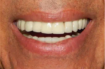
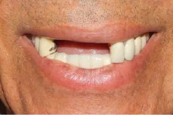
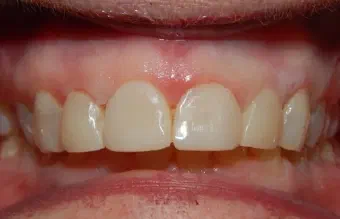
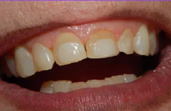
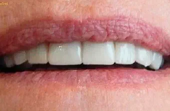
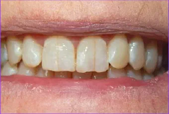
Before & after smile gallery.

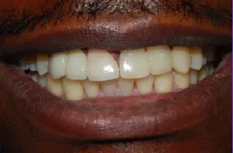
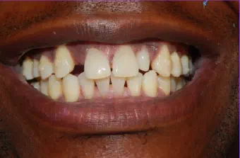
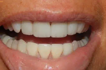
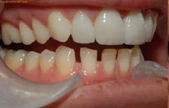
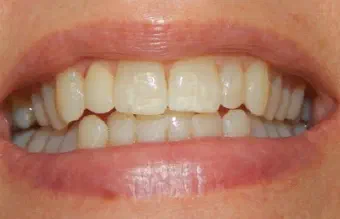
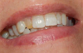
Drag the centre line to compare each real case treated by our dental team. Every smile begins with a personal assessment and a plan shaped around the patient.

Areas We Serve

Neighbourhood dental care, close to home.

Located in Cookstown, Innisfil, our dental team welcomes patients from communities across South Simcoe, Barrie, and northern York Region.

Get directions
  • Cookstown
  • Innisfil
  • Simcoe
  • Lake Simcoe
  • Alcona
  • Stroud
  • Lefroy–Belle Ewart
  • Gilford
  • Churchill
  • Sandy Cove
  • Innisfil Heights
  • Fennell’s Corners
  • Bradford
  • Bradford West Gwillimbury
  • Bond Head
  • Newton Robinson
  • New Tecumseth
  • Alliston
  • Beeton
  • Tottenham
  • Adjala–Tosorontio
  • Everett
  • Loretto
  • Barrie
  • Essa
  • Thornton
  • Baxter
  • Angus
  • Egbert
  • Ivy
  • Utopia
  • York Region
  • East Gwillimbury
  • Queensville
  • Holland Landing
  • Sharon
  • Schomberg
  • Nobleton

Don’t see your community listed? Contact us—we regularly welcome patients from throughout the surrounding region.

Google reviews

Kind words from our patients.

Read what patients have shared about their visits to Cookstown Dental Centre.

A dentist having a friendly consultation with a patient in a bright dental clinic

Your next step

Ready to feel good about your next dental visit?

Whether you are new to the clinic or returning for care, our team is here to make booking simple and answer your questions.

Call Book Now WhatsApp

How can we help?

The Cookstown Dental team is here for you.

Need an appointment, have a question, or want to talk through your options? Choose the next step that works best for you.

Send us a note

We’ll get back to you as soon as we can.