Cookstown Dental Centre
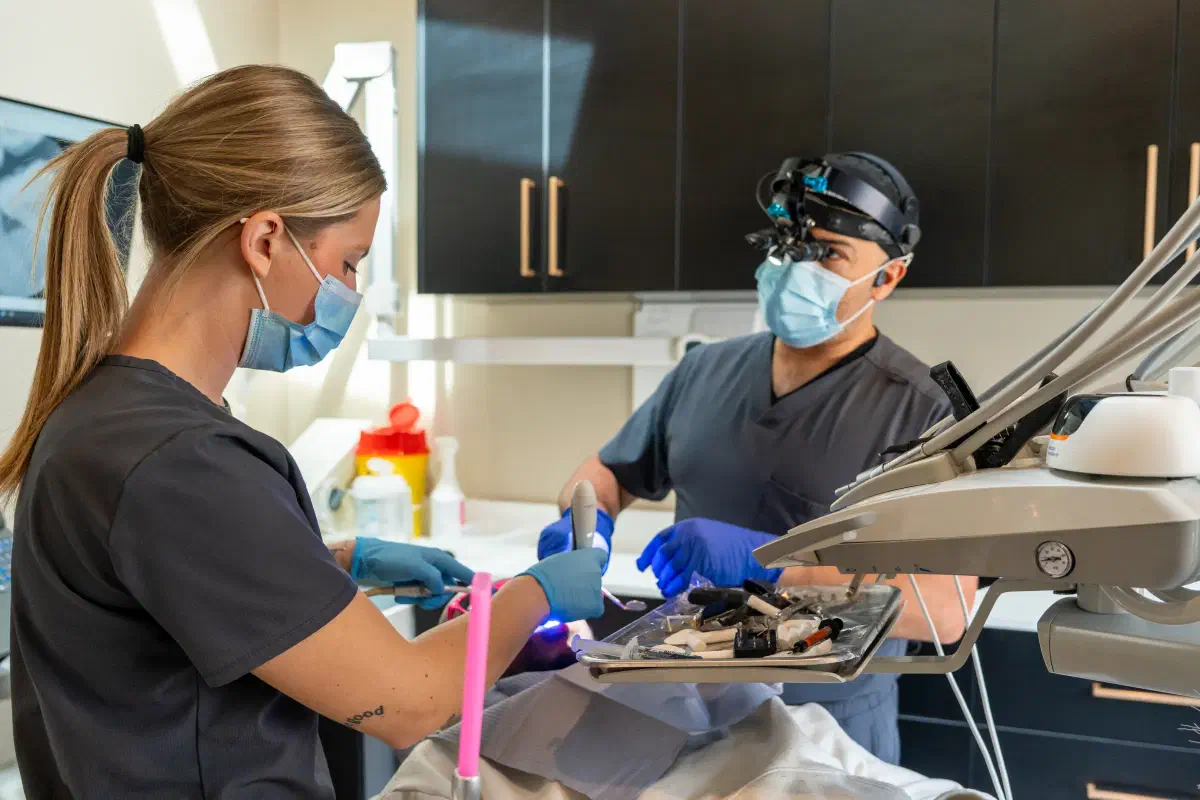
The Cookstown Dental Centre team caring for a patient in a treatment room

Specialized Care · Cookstown

Understand changes in the bone supporting your teeth.

Bone-loss assessment combines gum measurements, imaging, and treatment history. Clarify what has changed, whether disease is active, and how support affects care.

Request an appointment

Let’s find a time that works.

Need urgent dental help? Call (705) 291-2222.

Bone-Loss Assessment in Cookstown

An X-ray finding needs context before it becomes a treatment decision.

Bone around the teeth provides part of their supporting foundation. An assessment may be recommended when periodontal disease is suspected, a tooth becomes mobile, a radiograph shows reduced support, or treatment planning needs a clearer picture of the site. Bone changes around an implant or a healed extraction area require their own interpretation and should not be assumed identical to periodontal loss around natural teeth.

At Cookstown Dental Centre, we relate imaging to the clinical examination, your history, and previous records when available. One image can show the current level of support but may not establish when a change occurred or whether disease is progressing now. The purpose is to explain the findings, identify treatment priorities, and plan monitoring or referral without assuming that every area of reduced bone requires extraction or grafting.
A dentist explaining the bone supporting teeth with a jaw model during consultation

Good to know

Clarify which type of bone change is being assessed.

The location and history influence what the finding means.

Support around a natural tooth

Periodontal disease can affect attachment and supporting bone, while other local conditions may produce different patterns. The clinician examines the gums, roots, mobility, and related symptoms. The amount visible on an image is interpreted with these findings before discussing the tooth’s prognosis.

An implant site or missing-tooth ridge

Bone around an implant and ridge changes after extraction need an assessment suited to that situation. An implant-tissue concern may require peri-implant evaluation, while a deficient ridge may relate to future replacement planning. The term bone loss should not lead to one identical treatment for all locations.

What we consider

Combine measurements, images, and previous information.

Each contributes something different to the assessment of support.

Clinical periodontal findings

Pocket depths, bleeding, recession, attachment findings, and tooth mobility help describe the current condition. Measurements are interpreted together rather than used as isolated numbers. The dentist explains whether the pattern suggests active inflammation, a treated reduced-support situation, or another concern needing further evaluation.

Appropriate imaging

Radiographs can show aspects of bone support and local anatomy that cannot be seen directly in the mouth. The type and need for new imaging depend on the clinical question and existing records. A scan is not automatically required for every finding, and the provider should explain its purpose when recommended.

Comparison with earlier records

Previous measurements and images may help establish whether support has changed. Differences in image angle or record quality can affect interpretation, so comparisons require care. If older records are unavailable, a current baseline and planned review may help clarify the situation without pretending the timing of loss is already known.

Planning together

Consider the factors that affect prognosis and treatment planning.

The amount of remaining bone matters, but it is not the only determinant.

Inflammation, cleaning access, and risk factors

Deposits, home-care difficulties, smoking, diabetes, and periodontal history may influence the care plan. Discuss these without assuming that one factor explains every site. The goal is to identify manageable contributors and suitable monitoring rather than reduce the assessment to a single radiographic percentage.

Tooth structure, position, and function

A tooth’s root form, restorability, bite, and role in a planned prosthesis can affect its outlook. Reduced support does not automatically mean removal is necessary. The clinician should explain the combination of findings behind any recommendation to retain, treat, monitor, or remove a particular tooth.

Step by step

Leave with a baseline and a plan for review.

A useful assessment makes the uncertainty and next decisions visible.

  1. Understand the findings in plain language

    Ask which teeth or sites are affected, what evidence supports the diagnosis, and whether progression can be established. The provider should distinguish known findings from uncertainty. That explanation helps you understand why a treatment or review interval is recommended rather than reacting only to the phrase bone loss.

  2. Prioritize treatment and provider coordination

    Urgent infection, unstable teeth, and planned restorative work may affect sequencing. If a periodontist or another clinician is involved, clarify the purpose of referral and how records will be shared. A coordinated plan avoids designing a restoration before its supporting tissues have been appropriately assessed.

  3. Monitor response and report new changes

    Follow the agreed care and review schedule. New movement, swelling, drainage, or biting discomfort should prompt contact between visits. A previously documented reduced level of support does not make every future symptom expected or harmless, and a stable period still needs ongoing preventive attention.

Frequently asked questions

Questions about bone-loss assessment

Does bone loss on an X-ray mean my tooth must come out?

No. The remaining support, inflammation, root form, tooth condition, and function all influence prognosis. Some teeth with reduced support can be maintained with appropriate care. The dentist should explain the combined findings behind any extraction recommendation.

Can one X-ray show whether the loss is still progressing?

An image can show current support but may not establish when the change occurred. Previous records, clinical findings, and follow-up can help assess progression. The provider should distinguish evidence of reduced support from certainty about ongoing loss.

Will a deep cleaning grow all the bone back?

That should not be promised. Nonsurgical periodontal care aims to manage deposits and inflammation, with response assessed afterward. Selected defects may have other treatment options, but the potential for regeneration depends on the site and clinical plan.

Is this assessment the same for an implant?

Implants require evaluation of their own tissues, support, and restoration. Bone findings around an implant are not automatically interpreted like periodontal findings around a natural tooth. The clinician may recommend a focused peri-implant assessment and relevant comparison records.

Will I need a three-dimensional scan?

Not automatically. Imaging is chosen according to the question and existing information. Conventional radiographs may be appropriate for many periodontal assessments, while selected planning needs can justify additional imaging. Ask what a proposed scan is expected to clarify.

Can bone loss happen without pain?

Yes. Periodontal changes may be present without noticeable pain, which is why examination and appropriate records matter. Lack of discomfort does not establish healthy support, and new mobility or gum changes should be assessed even when eating remains comfortable.

What records should I bring?

Previous relevant radiographs, periodontal measurements, treatment details, and implant information if applicable can help. Also update your medical and medication history. Missing older records does not prevent an assessment, but it can limit certainty about when or how quickly a change occurred.

Real patient results

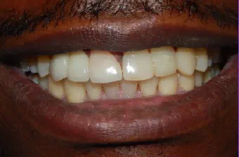
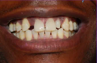
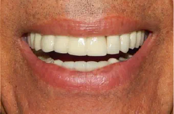
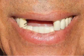
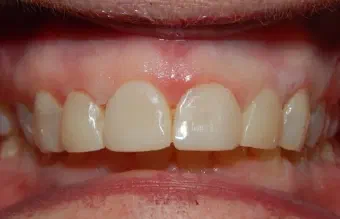
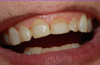
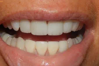
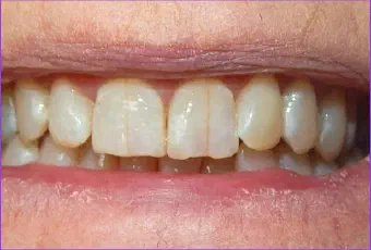
Before & after smile gallery.

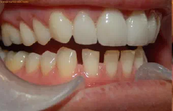
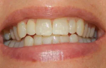
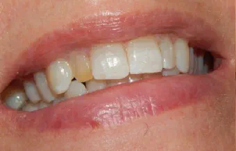
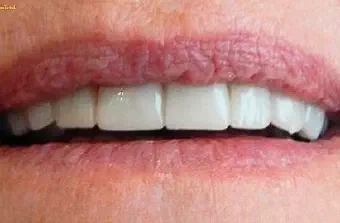
Drag the centre line to compare each real case treated by our dental team. Every smile begins with a personal assessment and a plan shaped around the patient.

Areas We Serve

Neighbourhood dental care, close to home.

Located in Cookstown, Innisfil, our dental team welcomes patients from communities across South Simcoe, Barrie, and northern York Region.

Get directions
  • Cookstown
  • Innisfil
  • Simcoe
  • Lake Simcoe
  • Alcona
  • Stroud
  • Lefroy–Belle Ewart
  • Gilford
  • Churchill
  • Sandy Cove
  • Innisfil Heights
  • Fennell’s Corners
  • Bradford
  • Bradford West Gwillimbury
  • Bond Head
  • Newton Robinson
  • New Tecumseth
  • Alliston
  • Beeton
  • Tottenham
  • Adjala–Tosorontio
  • Everett
  • Loretto
  • Barrie
  • Essa
  • Thornton
  • Baxter
  • Angus
  • Egbert
  • Ivy
  • Utopia
  • York Region
  • East Gwillimbury
  • Queensville
  • Holland Landing
  • Sharon
  • Schomberg
  • Nobleton

Don’t see your community listed? Contact us—we regularly welcome patients from throughout the surrounding region.

Google reviews

Kind words from our patients.

Read what patients have shared about their visits to Cookstown Dental Centre.

A dentist having a friendly consultation with a patient in a bright dental clinic

Your next step

Ready to feel good about your next dental visit?

Whether you are new to the clinic or returning for care, our team is here to make booking simple and answer your questions.

Call Book Now WhatsApp

How can we help?

The Cookstown Dental team is here for you.

Need an appointment, have a question, or want to talk through your options? Choose the next step that works best for you.

Send us a note

We’ll get back to you as soon as we can.